Noninvasive Cardiology
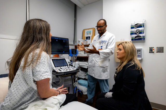
Noninvasive diagnostic testing and cardiovascular imaging at UF Health Jacksonville focuses on diagnosis and follow-up of heart disease by using state-of-the-art technologies. Our heart team experts may recommend a noninvasive cardiology test or procedure to evaluate or diagnose symptoms concerning of heart disease or to follow-up a preexisting heart condition.
Types of noninvasive cardiology procedures
There a number of noninvasive cardiology tests designed to help doctors diagnose a heart condition and determine the proper treatment. A team of experienced registered nurses, sonographers and technicians perform tests while attending to keeping patients comfortable and well informed during the test.
- Cardiac magnetic resonance imaging (MRI, CMRI) - Detailed pictures of the heart are taken by using radio-frequency waves and a strong magnetic field.
- Cardiac multi-slice computed tomography (CT) - During a CT scan of the heart and chest, a technician uses X-rays and a computer to capture detailed, three-dimensional images.
- Echocardiogram - A small device, called a transducer, is placed on a patient’s chest that emits ultrasound waves that produce detailed images of the heart. These images allow specialists to see the heart’s size, structure and motion to determine the health of the heart. Several types of echocardiogram procedures are available to aid cardiologists in determining the condition of the heart.
- Electrocardiogram (ECG) test records the electrical activity of the heart to help doctors determine rhythm abnormalities or to detect a prior heart attack.
- Event monitoring is a heart monitor worn for about 30 days to monitor heart rhythm and to record symptoms and is used to diagnose symptoms that are infrequent or sporadic.
- Holter monitoring is a heart monitor usually worn for 24 hours that continuously records a patient’s heart rhythm during typical daily activities. It is especially useful in diagnosing palpitations and detection of arrhythmia.
- Nuclear perfusion imaging is performed with exercise or with the use of medication to stress the heart in patients who cannot exercise. Very small amounts of radioactive tracers are injected into the bloodstream, and images of the heart muscle are obtained on a gamma camera, which helps in detection of coronary artery disease and in evaluation of heart function.
- Tilt table study determines the cause of fainting spells.
- Treadmill or cardiac stress test monitors the heart rhythm on EKG while a patient walks on a treadmill to evaluate symptoms such as chest pain, shortness of breath and fatigue; helps in diagnosis of arrhythmias during exercise; and may help in detection of any decrease in blood supply to the heart muscle due to blood vessel blockage (coronary artery disease).
- Vascular studies use ultrasound, or high-frequency sound waves, to examine the flow of blood in your vessels and detect vascular disease. There are several types of these noninvasive scans that can be done to help diagnose your condition.
Why choose UF Health for noninvasive cardiology?
At the UF Health Cardiovascular Center in Jacksonville, it is all about a patient’s heart and vascular health. Our goal is a thorough evaluation of symptoms by the use of state-of-the-art testing to accurately diagnose and follow-up any cardiac disease. To this purpose, the UF Health Cardiovascular Center, has invested in the most advanced diagnostic equipment to assist our team in determining the best possible treatment plan as quickly as possible. Our highly trained cardiologists, cardiovascular surgeons and staff provide compassionate and exceptional cardiac care for a variety of cardiovascular diseases.
Our locations
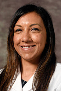
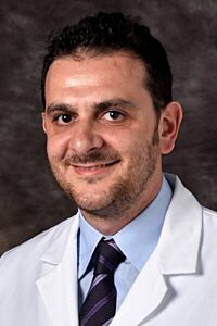
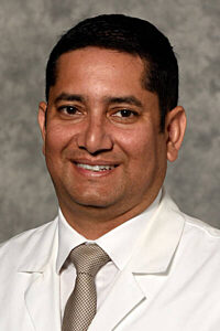
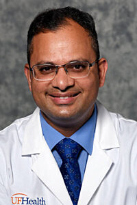
Jacksonville noninvasive cardiology specialists
-
Khadeeja Esmail, MDCardiologist

-

-
Dinesh Kadariya, MDCardiologist

-
Pankaj Mathur, MDCardiologist

-
Emil D Missov, MD, PhDCardiologist

-

-

-

-
Carlos R Zamora, MDCardiologist

