Arteriovenous malformation (AVM)
What is an arteriovenous malformation?
A brain or spine arteriovenous malformation, or AVM, is a tangle of arteries and veins that interferes with blood circulation. AVMs can occur anywhere, but they are more common in the brain or spinal cord. Many people have few symptoms until a rupture occurs; however, some people may experience symptoms depending on the location of the AVM.
Some indications of a brain AVM include seizure, weakness, numbness or headache, typically in one area of the head. Some symptoms may be severe. A spine AVM may cause weakness in the arms or legs, numbness and balance problems.

Arteriovenous malformation: Treatment
Treatment is based on the location, size and position of the draining vein associated with the AVM. Your physician will create a treatment plan specific to your needs to ensure care is given to tending to the delicate nature of this condition.
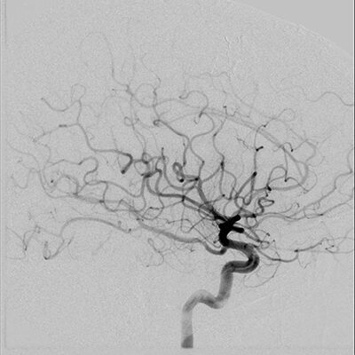
The first step in developing a treatment plan is to obtain a cerebral angiogram. Also known as digital subtraction angiography (DSA), this test uses a minimally invasive sterile procedure performed using fluoroscopy (live X-ray). A small tube called a catheter is used to access the femoral artery located in the groin or the radial artery at the wrist. The catheter is advanced over a guidewire to select vessels through which contrast is injected to obtain images.
During your visit, your physician will discuss your treatment options:
- Embolization is a common procedure used to block the blood flow of the AVM, which can make it easier to remove by surgery. A limited number of highly selected, small AVM can be treated with embolization only. The type of treatment depends on the location, size and location of feeding and draining vessels. Some of the AVM are observed over time with imaging rather than treated due to high risk of treatment or low lifetime risk of hemorrhage.
- Endovascular treatments are performed from within the vessel, typically accessed at the femoral artery located at the groin or radial artery at the wrist from which catheters are advanced over a small wire to the AVM. Again, the goal is to block the blood supply. Sometimes more than one treatment session is needed to block a majority or all the feeding vessels.
- Open surgical treatment, called a microsurgical resection, may be used either in conjunction with endovascular embolization or as the primary treatment. This option will be discussed at the time of your appointment.