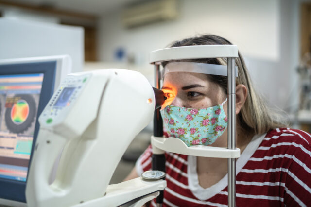
Could my blurry vision be cataracts?
People with a cataract in one or both eyes experience blurry vision from the clouding of the eye’s natural lens

Update your location to show providers, locations, and services closest to you.
A cataract is a clouding of the lens of the eye. It is a natural process and happens as we age. Most people will develop some form of cataract as they grow older, but there are many variables which can hasten cataract development. Common risk factors for earlier cataracts include family history, nutrition, sunlight exposure, prior eye surgery or diseases, eye trauma and systemic diseases such as diabetes. If a cataract is present, the decision to proceed with cataract surgery depends on both the ophthalmology exam and the patient’s visual needs.
Cataract surgery technologically advanced areas of ophthalmologic surgery. UF Health Ophthalmology – Jacksonville offers advanced cataract surgery techniques in its ambulatory surgery center. Several qualified, board-certified ophthalmologists routinely perform this procedure.
Some common forms of cataract include:
Patients with cataracts will usually note decreased, blurry or even dull vision. They may experience glare from lights or loss of color saturation.
The diagnosis of a cataract is typically done with a slit-lamp exam. The eye is examined with a special microscope to evaluate any changes in the natural clarity of the lens. It is important that the entire eye is examined, including the retina, to make sure that there are no other diseases or abnormalities that might be causing visual problems.
In some circumstances, additional tests are done to evaluate the vision in a patient affected by a cataract. These tests include:
Patients with a cataract should undergo a complete eye exam to rule out other causes of visual loss, such as macular degeneration, a disorder that affects the central part of the retina of the eye causing decreased vision and possible loss of central vision, or glaucoma, a disease of the nerve that carries visual information from the eye to the brain. If the primary cause of vision loss is determined to be the cataract, treatment options are limited and include observation, wearing new glasses to improve vision or surgery. There are no eye drops or nutritional supplements that are known to remove cataracts. In addition to decreased visual acuity, patients sometimes choose to have cataract surgery because of problems with glare, loss of contrast sensitivity, changes in the focusing power of the eye or double vision.
Cataract surgery is routinely performed with the patient awake. Special medications are placed behind the eye with a nerve block or topical solution on the eye. These medications numb the eye so that the surgeon can make a small incision and carefully remove the cataract. While ultrasound vibrations generally are used to remove cataracts, there are some cases in which the cataract is removed manually. Once removed, a new lens made of special plastic material can be placed in the eye. The patient is usually home the same day.
Cataract surgery does not typically hurt, as most surgery is performed under topical anesthesia. This means the patient is awake and the eye is numb. The patient may feel some pressure and movements, but does not experience sharp pain. If the patient does experience discomfort, the physician can immediately add more numbing medications.
The whole process of cataract surgery from beginning to end takes about half a day. This includes check in, pre-operative assessment, surgery and post-operative care. The surgery takes about 30 minutes.
Cataract surgery is generally considered to be a relatively safe and effective surgery. However, like any surgery, there are unique risks. More common risks include infection, glaucoma, inflammation, retinal detachment and the inability to place an intraocular lens at the time of the initial surgery.
After cataract surgery, patients usually notice improvement in their vision within days. However, due to the delicate nature of eye surgery, patients are asked to take certain medications and restrict strenuous activities for several weeks.


People with a cataract in one or both eyes experience blurry vision from the clouding of the eye’s natural lens

We continue our celebration of Asian American and Pacific Islander, or AAPI, Heritage Month, by featuring Darrell WuDunn, MD.
