- Investigator
- Mobeen H Rathore
- Status
- Accepting Candidates
- Ages
- 3 Years - 6 Years
- Sexes
- All
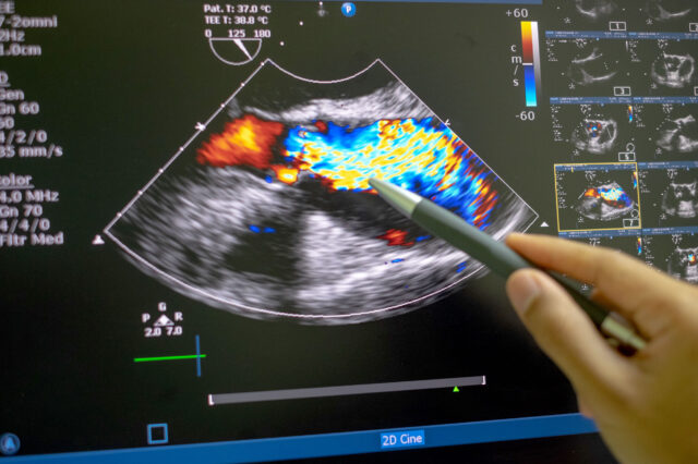
During an echocardiogram, a small device called a transducer is placed on a patient’s chest. Using ultrasound waves, the device produces detailed images of the heart. These images allow specialists to see the heart’s size, structure and motion to determine the health of the heart.
Several types of echocardiogram procedures are available to aid cardiologists in determining the condition of the heart.
- Exercise stress echocardiogram is used to detect and evaluate symptoms of chest pain or shortness of breath, in detection of coronary artery disease, while pedaling a stationary bike or exercising on a treadmill. The echo images obtained at rest and various stages of exercise help doctors to understand the heart function, in detection of any CAD and to assess a patient’s exercise capacity and symptoms.
- Dobutamine stress echocardiogram is a noninvasive stress test reserved for individuals unable to exercise. It detects and evaluates coronary artery disease during rest and when the heart is beating fast. A medication called Dobutamine is administered through an intravenous line to raise the heart rate to the level required with constant monitoring of the heart on EKG and getting serial echocardiographic images to understand the heart response to stress.
- Two-dimensional and three-dimensional transthoracic echocardiogram (TTE) use ultrasound waves to create an image of the heart to evaluate the structure and functioning of the heart for diagnosis of cardiac disease and abnormalities. The 3D echo visualizes the heart in real time in stunning detail to precisely locate abnormalities in complex diseases.
- Transesophageal echocardiogram (TEE) and three-dimensional TEE are used when accurate images of the heart chambers and valves cannot be imaged satisfactorily from outside the chest wall as in a transthoracic echocardiogram. Transesophageal echocardiography is an ultrasound imaging exam where, after a topical numbing medicine and a sedative are administered, an imaging probe is introduced into the esophagus like in endoscopy. Live images are taken during the study to assess the heart and its chambers, valves and major blood vessels, giving a much clearer view of those structures than other imaging methods.
Related conditions & treatments
Clinical Trials: Echocardiogram
UF Health research scientists make medicine better every day. They discover new ways to help people by running clinical trials. When you join a clinical trial, you can get advanced medical care. Sometimes years before it's available everywhere. You can also help make medicine better for everyone else. If you'd like to learn more about clinical trials, visit our clinical trials page. Or click one of the links below:
News and Patient Stories: Echocardiogram
Getting a second chance at life
How the minimally invasive MitraClip procedure helped Karen Kaunitz's battle with lung disease.

Saving the heart that is saving the world
July 31, 2018
They were once considered the mythical unicorns of the forest, with many people unsure they even existed. The large mammal looks like a mix of deer, giraffe…


